10
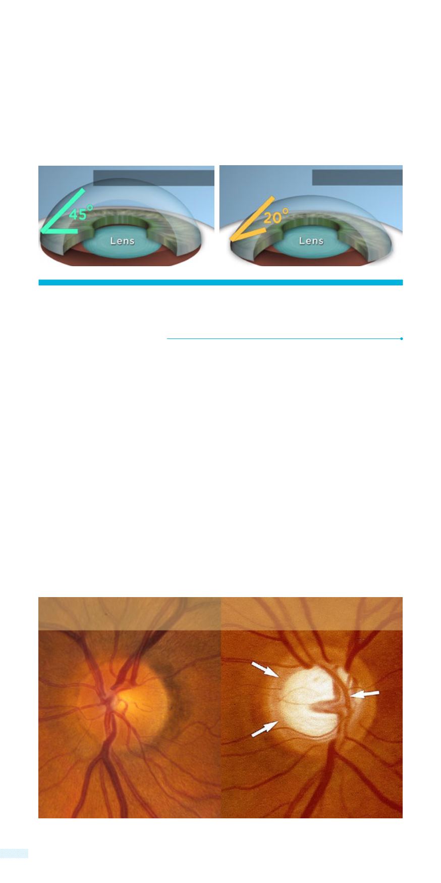
Even before glaucoma reveals its first symptoms, it is possible for
the ophthalmologist to discover morphological changes of the
optic disc with a simple fundoscopy. This is usually performed with
the installation of mydriatic drops so that the fundus can be seen
through the dilated pupil.
Thinning of the nerve fiber layer and increase of the normal cupping
of the optic disc, as well as disturbances in the path of the small
blood vessels and/or small hemorrhages, are some of the findings
that an ophthalmologist can take into consideration in order to
determine the range of damage.
Fundoscopic evaluation
of the optic disc
calibration systems that measure the width of the angle, but generally
we use the term “narrow angle” to describe a small distance between
the iris and the interior surface of the cornea which anatomically
restrict the drainage of the aqueous and leads to a rise in intraocular
pressure.
Normal Angle
Narrow Angle
OPTIC DISC
Normal
Glaucoma


