17
The international practice for the treatment of glaucoma aims at
the reduction of intraocular pressure, and the objective is to avoid
further damage to the optic nerve. The target pressure however is
not the same for everyone.
In a patient with early glaucoma, a reduction to 17-18mmHg will
probably suffice to inhibit disease progression. In a patient with
advanced disease the safe levels are much lower, even at about 10-
12mmHg. In every case, the calculation of the optimal pressure and
the way it is achieved, have to be personalized.
Although the modern anti-glaucoma drugs are very effective,
especially in Primary Open Angle Glaucoma, some patients might
not be regulated correctly and then other secondary treatments will
be needed (Laser and/or surgery).
At Athens Eye Hospital, anti
glaucomatous treatment is
adapted to each patient’s needs
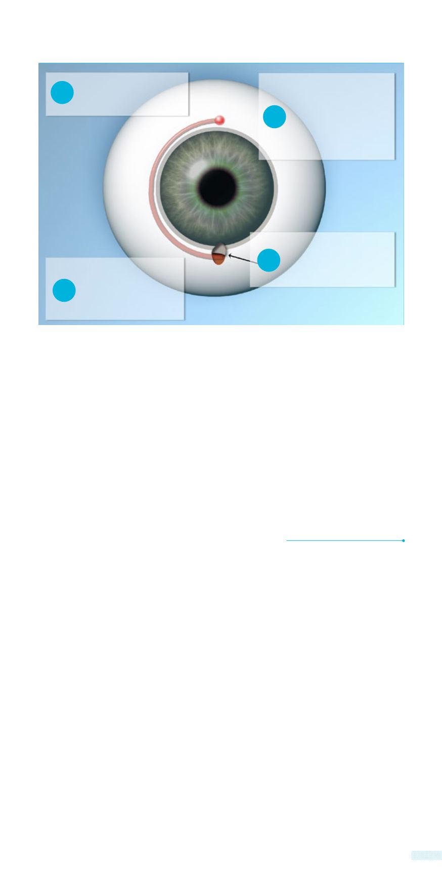
This catheter has
a guidance light at
one end. Within its
tube, a special gel is
injected that causes
expansion of the
drainage system
A tiny catheter is
forwarded from a micro-
incision around the
drainage canal of the eye.
CANALOPLASTY
Light
A thin suture follows the
micro catheter in the
canal all around 360
o
in the periphery
The catheter is removed
and the suture is
tightened so that the
drainage canal stretches
internally and remains
permanently open.
1
2
3
4
Incision
system causing its dilation. A very thin suture follows the micro
catheter in its circular direction in the channel. After removing of the
catheter, the suture tightens to stretch the channel and keep it open
permanently.


