7
Even before injecting any dye, the fundus
fluorescences
due to a
natural dye called “lipofuscin”. This natural autofluorescence provides
us with vital information about the condition of the
Retinal Pigment
Epithelium
(RPE), the layer of cells that provides nutrients for the
retina.
In many conditions including ARMD, the RPE is dysfunctioning and
cannot displace enough the lipofuscin, resulting in accumulation and
autofluorescence.
The angiography department of Athens Eye Hospital is
on call 24 hours with a specialist ophthalmologist
These are special photographic techniques of the fundus, where
special dyes (fluorescein or indocyanine) are injected intravenously.
In
Athens Eye Hospital
we have the most up-to-date imaging
systems as the SPECTRALIS® HRA, which takes advantage of the
accuracy of a special laser for even better sharpness and depth in the
images, but is also able to perform parallel angiographs (fluorescein
& indocyanine) thus reducing the time needed.
These examinations can yield key information on the condition of
the arteries and the veins that lie in the internal eye, and hence are
of vital diagnostic importance for ARMD as well as numerous other
conditions.
Fundus Autofluorescence (FAF)
Fluoroangiography and
Indocyanine green angiography
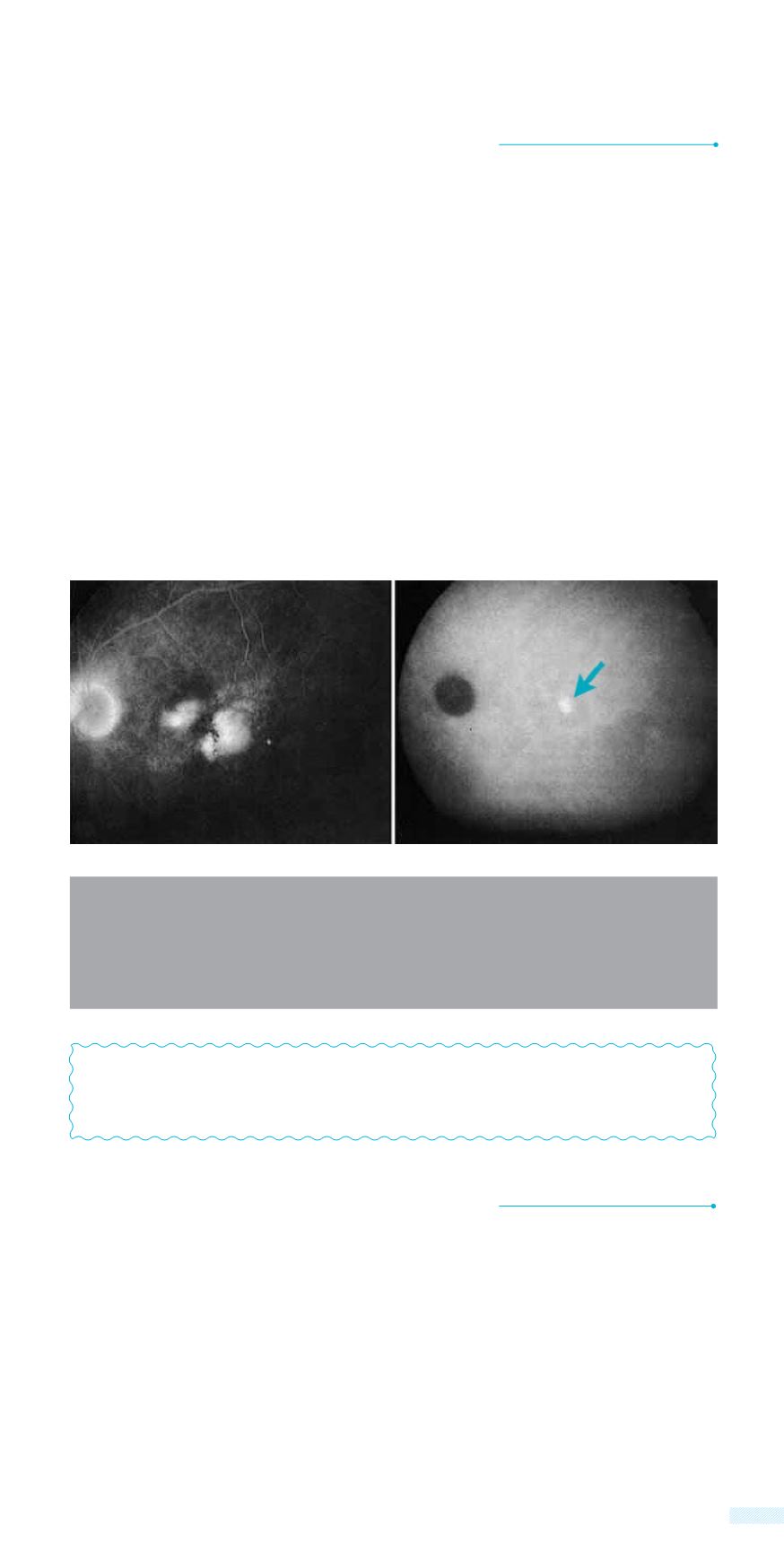
A: Late phase fluoroangiography showing leakage from the neovessels in a
patient with wet ARMD
B: Late phase indocyanine green angiography on the same patient,
showing the exact spot of leakage
Α
B


