8
The OCT is an imaging system that allows the analysis of the retina in
the macular area, depicting transverse segments. The examination is
painless and very fast, without utilizing radiation in any form, dilating
drops or the need for injecting any dye.
An OCT examination can reveal even the smallest changes or lesions
in the retinal structure, evaluate and monitor the progression of the
disease in the macula, as well as the correspondence to therapy.
The “Time Domain OCT” (TD-OCT) were released first, with a reso-
lution of 10μm (one-millionth of a meter). Later came the “Spectral
Domain OCT” (SD-OCT), improving the resolution to 1-3μm, with a
40,000 scans/sec capability, and also added the ability of a 3-Di-
mensional depiction of the retina.
Athens Eye Hospital
possesses all the available equipment to record
autofluorescence, that in conjunction with digital angiography with
indocyanine, can contribute in
occult neovascularization
detection
as well as in the differential diagnosis between “Idiopathic Polypoidal
Choroidal Vasculopathy” (IPCV) and “Retinal Angiomatous Prolifer-
ation” (RAP). The distinction between all these conditions is of the
utmost importance since they correspond differently to therapy.
Optical Coherence Tomography (OCT)
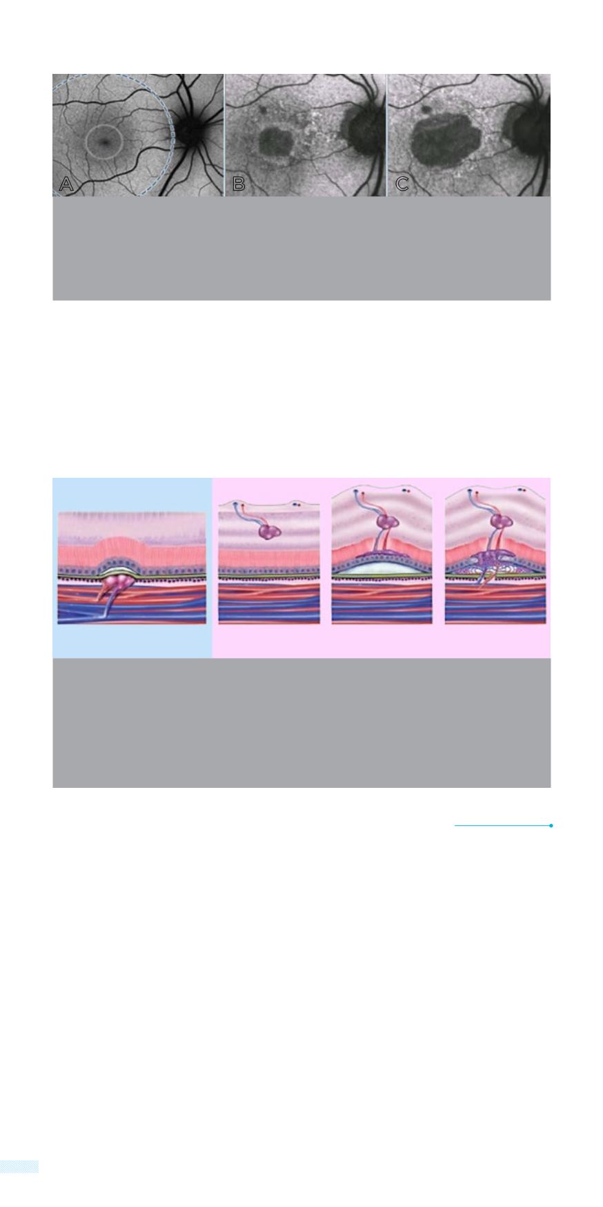
Autofluorescence imaging with a special blue laser (Blue peak Blue Laser
autofluorescence)
Figure A: Normal
Figure B: Geographic atrophy with increased fundus autofluorescence
Figure C: Expansion of the atrophy on the same patient 3 years later
In Idiopathic Polypoidal Choroidal Vasculopathy (IPCV), neovascular-
ization takes place at the choroid where polypoidal arrangements are
forming. In Retinal Angiomatous Proliferation” (RAP), new vessels ini-
tiate from the retina and then invade the choroid. Distinction between
these forms of neovascularization is very important as they have differ-
ent prognosis and require a different therapeutic approach.
ON
ON
ON
C
Β
Α
IPCV
RAP


