13
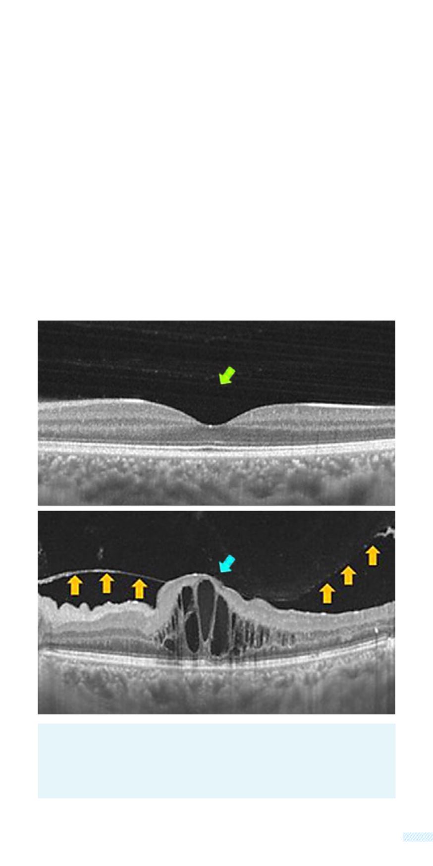
Swept Source OCT:
Cyctoid macula edema (blue arrow). High
accumulation of fluid in the area of the central fovea organized in
cystic formation. The yellow arrow show the vitreous which has been
detached. In comparison to the normal appearance (green arrow).
Normal
Cystoid Macula Edema
An OCT examination can reveal the smallest changes or deterioration
in the structure of the retina. It is totally necessary in the initial
evaluation of macular edema and the monitoring of the therapeutic
outcome.
The first generation were Time Domain OCT (TD OCT) machines,
with resolution of 10 (ten) μm (microns - millionth of a meter). The
next evolution were the Spectral Domain OCT (SD OCT), improving
the resolution to 1-3μmwith 40000 A Scans per second, accelerating
examination speed faster and adding a 3-dimentional capability, in
the mapping of the retina.
At
Athens Eye Hospital
we have the latest generation Swept Source
OCT, with the capability of 100000 A-scans per second and a greater
penetrating capability for better control.


